napari Tutorial DL at MBL 2024
This is the napari tutorial for DL at MBL 2024
Aug 27, 2024
Polarity-JaM: An image analysis toolbox for cell polarity, junction and morphology quantification
Polarity-JaM is an open-source Python package for image analysis of cell polarity, junction, and morphology, featuring deep learning-based segmentation and advanced statistical tools for comprehensive analysis.
Apr 26, 2024
RS-FISH: precise, interactive, fast, and scalable FISH spot detection
RS-FISH is a software tool for accurate and fast spot detection in microscopy images, supporting large datasets and offering interactive parameter tuning.
Dec 1, 2022
napari
napari is a Python viewer for multi-dimensional images. I joined CZI’s napari team in 2022, where I made code contributions and served as tech lead. I’ve been on napari’s steering council since 2023. napari has become central infrastructure in the scientific Python ecosystem for microscopy—used for everything from basic image visualization to interactive ML annotation workflows. My contributions have focused on large-data handling, ML integration, and helping coordinate the project’s technical direction.
Jul 1, 2022
Algorithms and computational tools for image data analysis across scales (keynote)
Keynote on computational tools for multi-scale image analysis.
Sep 1, 2021
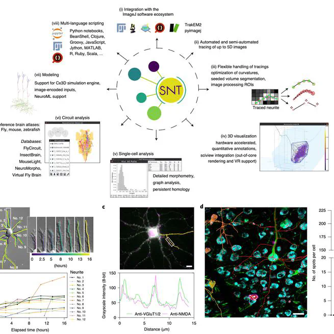
SNT: a unifying toolbox for quantification of neuronal anatomy
SNT is a comprehensive framework for neuronal morphometry and connectomics, providing tools for tracing, proof-editing, visualization, quantification, and modeling of neuroanatomy, available via Fiji's ImageJ distribution.
Apr 1, 2021
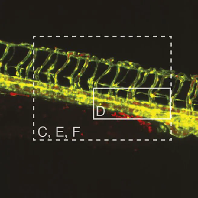
Multi-sample SPIM image acquisition, processing and analysis of vascular growth in zebrafish
We present a workflow for multi-sample SPIM imaging, processing, and analysis to study vascular growth in zebrafish, allowing simultaneous imaging and automated data processing, yielding precise quantification of vascular development.
Mar 20, 2019