Multi-sample SPIM image acquisition, processing and analysis of vascular growth in zebrafish
Mar 20, 2019·,,,,·
0 min read
Stephan Daetwyler
Ulrik Günther
Carl D. Modes
Kyle I. S. Harrington
Jan Huisken
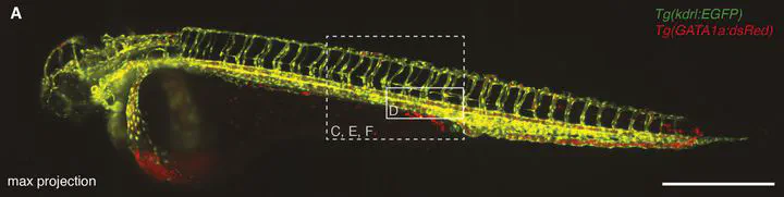 Image credit:
Abstract
To quantitatively understand biological processes that occur over many hours or days, it is desirable to image multiple samples simultaneously, and automatically process and analyse the resulting datasets. Here, we present a complete multi-sample preparation, imaging, processing and analysis workflow to determine the development of the vascular volume in zebrafish. Up to five live embryos were mounted and imaged simultaneously over several days using selective plane illumination microscopy (SPIM). The resulting large imagery dataset of several terabytes was processed in an automated manner on a high-performance computer cluster and segmented using a novel segmentation approach that uses images of red blood cells as training data. This analysis yielded a precise quantification of growth characteristics of the whole vascular network, head vasculature and tail vasculature over development. Our multi-sample platform demonstrates effective upgrades to conventional single-sample imaging platforms and paves the way for diverse quantitative long-term imaging studies.
Type
Publication
Development, 146(dev173757)